This is how our application simplifies the booking and administration process. Interested >>
Cruciate ligament surgery
A surgical intervention may become necessary, if the complaints indicate the damage of the anterior cruciate ligament (ACL), which is one of the most important ligaments responsible for knee joint stability.

Anterior cruciate ligament (ACL)
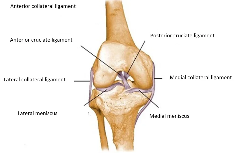
The knee joint has one of the most complex structures in the human body. The knee joint is involved in numerous movements of the lower limb, which is due to its location. The active (provided by muscles) and passive (provided by ligaments, bones, and cartilages) stability of the joint is essential to perform movements smoothly. The anterior cruciate ligament is one of the stabilizers of the knee joint, which is located within it, in between the thighbone, and the shinbone. Given its anatomical position, the anterior cruciate ligament inhibits the lower leg from sliding forward and back, and also from the internal rotation of the lower leg bones. Nerve endings that sense position and balance (proprioceptors) are present in the fibers of the anterior cruciate ligament, and they provide important feedback to the body; therefore, they are extremely important in regulating movement coordination. Instability develops in the knee joint if these functions are lost, and this causes difficulties both in the activities of daily living, and in sport as well. Additionally, since an unstable knee does not only roll, but also slides, the wear of the knee joint is accelerated in the long term.
General information about the rupture of the anterior cruciate ligament, and knee joint instability
The anterior cruciate ligament is most commonly injured by intensive rotational forces affecting the knee. These types of injuries are commonly associated with sport activities such as football, handball, basketball, skiing, martial arts, and dancing. The number of knee injuries is increased by the growing popularity of recreational sports, among which the injury of the anterior cruciate ligament is the most common. Typically, it occurs when the knee is exposed to stress coming from the side (valgus), overstretching (hyperextension), and external rotation and twisting. The ligament injuries of the knee joint occur mainly due to those movements, which exceed the joint’s normal range of motion. Complex injuries commonly occur during a ligament injury, so different structures may get injured at the same time within the joint, such as knee cartilages (menisci), cartilage surface of the joint, and other ligaments. The instability of the joint may pose risk for additional injuries in the future; therefore, the aim of our treatment is to restore a level of joint stability, which is consistent with the original anatomical set-up. This reduces the load affecting the knee joint surfaces. The surgical intervention provides solution for these injuries at the same time.
Which are the potential symptoms of an anterior cruciate ligament rupture, and knee joint instability?
The following are the most common symptoms of an anterior cruciate ligament rupture, which may occur with other knee joint injuries:
- Swelling
- Instability
- Pain
- Reduced range of motion
Specific symptoms of an anterior cruciate ligament rupture are the following:
- Knee joint instability
- Popping sound (when the fibrous ligaments tear).
What treatment options are available?
Basically, there are two types of treatment options available. Non-surgical (conservative), and surgical treatment.
The aim of conservative treatment
The main goal of the treatments is to achieve a painless condition, restore the range of motion and the stability of the knee. Unfortunately, the torn tendon does not heal with conservative treatment, so it is quite common that the pain, the knee joint instability, and temporal swellings do not resolve. It should also be expected that the joint wear of the knee will increase with time, which in years, may result in a knee joint that is painful and difficult to use even in everyday life. Therefore, surgical reconstruction is recommended in young age, if possible, instead of conservative treatment; the anterior cruciate ligament is replaced with the patient’s own tendon.
Conservative treatment methods
Physiotherapy, and other methods of physical therapy: PNF technique, kinematic taping, cryotherapy. Using ice is important to prevent inflammation, and to stop fluid retention in the joint. Stabilizers are strengthened by physiotherapy, muscle strengthening exercises, and PNF technique, and this provides acceptable stability until the surgery is performed.
Surgical treatment
Tendons from other parts of the body are used during the reconstruction of the anterior cruciate ligament. The implantable graft is most commonly prepared by using the hip flexor muscles. The patellar tendon is also used frequently. 7-10 days of unloading is necessary after the intervention. In non-problematic cases, the patients are discharged home 2-3 days after the surgery. The reconstructive surgery is most commonly performed in spinal anesthesia, and more rarely in general anesthesia, in a bloodless state, which is started from the root of the thigh. Antibiotic and anti-pathological coagulation prophylaxis are given during the surgery. It is necessary to regularly perform the practiced physiotherapy exercises at home, and to unload the limb with aids. After surgery, anticoagulants should be used for a defined period of time, which could be administered by the patients themselves. It can occur in certain cases that this type of reconstruction cannot be performed, or only partially. In a case like that, a different surgical solution or conservative therapy should be selected. The patient’s muscle strength may temporarily decrease with the intervention, but usually this is not significant from the perspective of an everyday lifestyle.
What happens during knee ligament surgery?

The surgical process
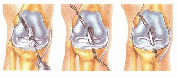
First, the location of the newly implantable ligament is cleansed during an arthroscopic intervention; the inflamed tissues are removed, and any potential knee cartilage (meniscus) and surface injuries of the cartilage are treated. Following this, the previously mentioned tendons are removed. For the purpose of graft removal, a skin incision of 3-4 cm is made during surgery at the inner side of the lower leg if the tendons of the knee flexors are used, and an incision of 7-8 cm below the patella if the patellar tendon is used.
After this, a hole is drilled in the bone structure of the thighbone and the shinbone, then the removed tendons are pulled in by an arthroscopic method. These are secured with a metal plate (endobutton) to the thighbone and with staples to the shinbone. (Figure 2)
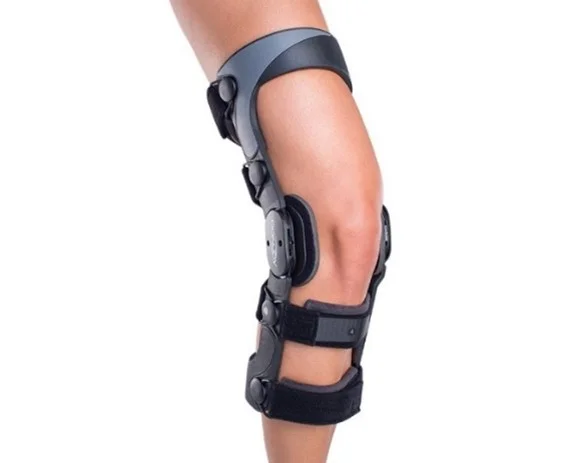
If the patellar tendon is used, a screw-driven or the so-called press-fit fixation is used. The area is rinsed, so that the inflammatory fluid and the debris are removed. If necessary, a small suction drain is placed into the wound, which drains the blood and serum that accumulate after surgery. The wound is closed (sewed) in multiple layers, and it is covered by sterile dressing. An external brace is applied after the surgery.

What happens if the justified surgical treatment is not performed?
Instability may increase, because of which doing sports may become unfeasible, and the cartilage surface may get damaged due to the instability of the knee joint, which may lead to joint wear in the long term (arthrosis). This process is irreversible, and it may result in a very painful joint with reduced range of motion, which often can only be treated by surgery. The pain may increase without treatment, and the instability may hinder everyday movements too.
If you have any questions, please send a letter to magankorhaz@bhc.hu!


