This is how our application simplifies the booking and administration process. Interested >>
Knee joint debridement
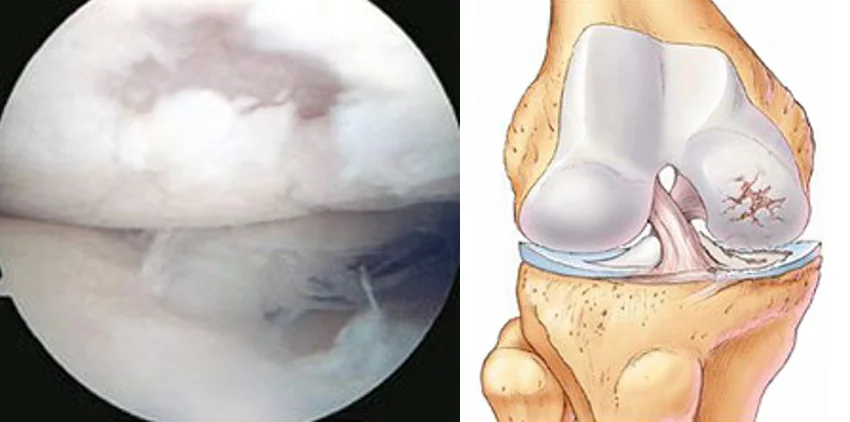
An arthroscopic intervention (knee joint debridement) may become necessary if the damage of the hyaline cartilage, which covers the bone surfaces (thighbone, shinbone), cannot be effectively treated with conservative methods.

What is the hyaline cartilage of the knee joint?
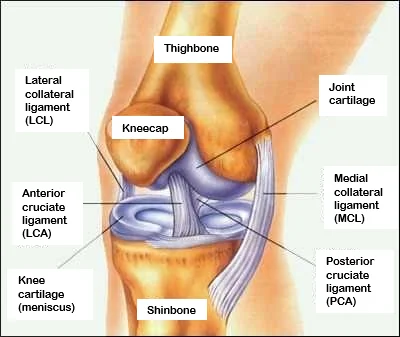
The knee joint has one of the most complex structures in the human body. The structure of the knee joint allows it to contribute to multiple lower limb movements. The surface of the thighbone and the shinbone are covered with hyaline cartilage, which is a specifically resilient tissue; therefore, it is mainly found in those areas of the body which are exposed to compression, but overload may damage the cartilage surface of the joint and induce pain in the knee joint. The bone surfaces directly touch if they are not adequately covered with cartilage, which may be associated with severe pain, and this could induce an inflammation in the joint.
General information about the disease
Cartilage surface damage of the knee joint mainly affects older patients, but it occurs in young athletes too, because the main cause of the damage is the constant overload of the cartilage surface. Additionally, potential knee injuries may also lead to cartilage surface damage. The resilience of the knee joint may decrease as a consequence of cartilage damage, and the range of motion may be reduced due to the pain. The extent of the damage may vary, and the decision about the necessity of potential interventions will be made depending on this. Knee instability or the axis deviation of the knee (X leg, O leg) could cause overload in the cartilage surface in numerous cases, which is the result of uneven loading. If the damaged cartilage is not treated in time, fragments may detach from the cartilage surface, and these could wear down the joint surfaces. This may lead to joint wear (arthrosis) over time. The goal of the surgery is to protect the cartilage surfaces of the knee joint from potential additional damage, and to prevent joint wear that develops over time.
What are the potential symptoms?
The most common symptoms of cartilage surface damage are the following:
- Pain
- Swelling
- Obstruction
- Reduced range of motion
- Pain of initiation (pain at the start of the movement, especially during the first few steps)
- Alternating periods with pain and without pain

What treatment options are available?
Basically, there are two treatment options available. Conservative (non surgical) and surgical treatment.
The aim of conservative treatment
The main goal of the treatments is to achieve a painless condition and to restore the knee’s range of motion. Unfortunately, the damaged cartilage surface does not heal with conservative treatment, so it is quite common that the pain and the knee’s reduced range of motion persist. It should also be expected that the joint wear of the knee will increase with time, which in years, may result in a knee joint that is painful and difficult to use even in everyday life.
Conservative treatment methods
It is necessary to elevate, and cool (icing, gel ice packs) the injured limb, if the knee is obstructed. Generally, it is not recommended to continue with sport. Fluid retention may develop as a consequence of the damage, and the knee may swell up. Conservative treatment includes pharmacological treatment, using painkillers and anti inflammatory drugs, physiotherapy, and other methods of physical therapy.
Surgical treatment
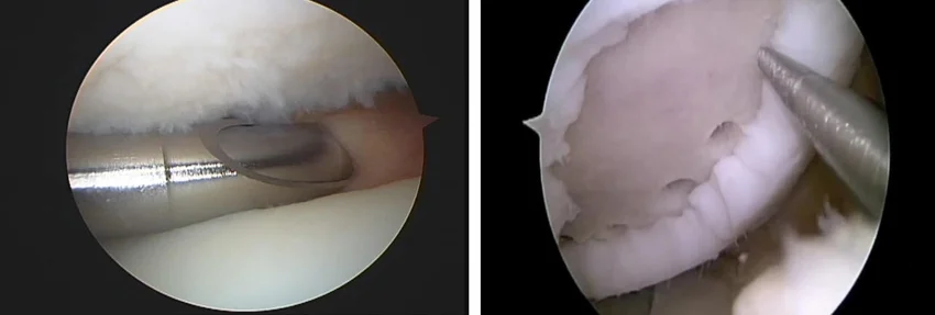
Treatment of hyaline cartilage damage is performed small incisions with a minimal invasive, arthroscopic intervention. The advantage of an arthroscopic intervention is that it is less demanding than an open surgery, so the pain after the surgery is less intense, and the duration of recovery is shorter too. It is ideal to perform the surgery as soon as possible after the injury, if it is associated with obstructions and swelling, so that the cartilage surfaces could be protected from further damage. Most commonly, the surgery is performed in general anesthesia (narcosis). The aim of the arthroscopic intervention is to remove the damaged, and detached cartilage fragments by sparing as much hyaline cartilage as possible. Following this, the remaining uneven cartilage surface is shaved down. Once the damaged bits are removed, the surface of the cartilage layer will become smaller, but it will still be able to perform basic functions, unless the extent of the damage is not too large.
Only two to three small cuts will be visible on the knee after the arthroscopic surgery. Recovery heavily relies on the type of surgery, and the postoperative treatment in which physiotherapists provide help. Antibiotic and pathological coagulation prophylaxis is used during surgery, if necessary. It is necessary to perform the practiced physiotherapy exercises, and to unload the operated limb at home.
An unloading period of three weeks is necessary after the intervention. In non problematic cases, the patients are discharged home on the first day after surgery. It may become necessary to apply thrombosis prophylaxis for a defined period of time after the surgery, and this could also be administered by the patient. It could happen during surgery that an extensive cartilage surface damage is revealed, which involves the bone too; this may necessitate further surgeries.
In this case, small drillings, and superficial microfractures are created beneath the cartilage surface (microfracture surgery), which may induce fibrous cartilage production in the areas with cartilage loss. Although, this is neither histologically, nor biomechanically identical to the original hyaline cartilage, which has a higher loading capacity. However, it is suitable to fill up the areas which are lacking cartilage and provide some level of load bearing.
What happens to you in the operating room?
The surgical process
During the surgery, two to three incisions of 1 cm are made on both sides of the knee, so that the arthroscopic optics and devices could be inserted into the knee joint. Special manual devices and a motor shaver are used during the intervention to remove the areas of damaged cartilage, and the inflamed tissue fragments. If the cartilage surface damage is well defined, and its extent is not large, then microfractures are created on the joint surface, which support the healing effect of the adjacent cartilage cells. Unfortunately, the fibrous cartilage which is produced this way is not consistent with the previous hyaline cartilage, but this intervention often provides a painless condition for the patients. The area is rinsed, so that the inflammatory fluid and the debris are removed. If necessary, a small suction drain is placed into the wound, which drains the blood and serum after surgery.
Elastic bandage is applied on the patient’s knee after the surgery.

What happens if the justified surgical treatment is not performed?
The damage of the cartilage surfaces may continue, which makes the pain and the feeling of obstruction more intense in the knee joint. The loose cartilage pieces may further damage the cartilage surface, which may lead to joint wear (arthrosis) over time. This process is irreversible, and it may result in a very painful joint with reduced range of motion, which could only be managed by surgery.
When can you return to your everyday activities?
Sedentary work usually can be started 1 3 weeks after surgery, but physical labor can only be started 6 weeks after the intervention. If microfractures were also created during the surgery, sometimes this period could be 2 3 months. When it comes to sports, the patient can start using a stationary bike after 5 10 days, swimming could be started after 2 3 weeks and jogging can be started after 3 4 weeks provided that the wound has healed. In order to achieve full recovery, other sports (football, basketball, etc.) can only be started 4 6 weeks after the intervention. If a surgery with microfractures was performed, riding a bicycle can be started without resistance after 7 10 days. Swimming can be started after the wound has healed, but other sports which load the lower limbs more can be only started 3 4 months after the surgery.
If you have any questions, please send a letter to magankorhaz@bhc.hu!


