This is how our application simplifies the booking and administration process. Interested >>
Rotator cuff reconstruction
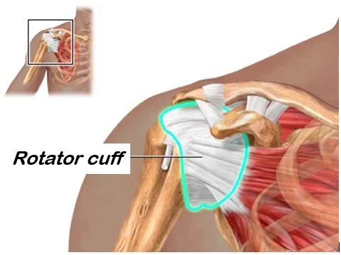
The rotator cuff is a group of shoulder muscles and tendons, which secures the upper arm bone to the shoulder joint. Rotator cuff diseases mainly affect people in the age of 50-60 years, but other than age, there are additional predisposing factors. Surgery may become necessary if the non surgical treatments are not effective.
What is the rotator cuff?
The rotator cuff consists of four muscles, which surround and move the head of the upper arm bone to multiple directions. The function of the muscle cuff is to make sure that the upper arm moves along with even forces, so that the head of the upper arm bone could roll in the joint socket in the most optimal position. The injury most commonly affects the supraspinatus muscle, which is located above the spine of the shoulder blade. This muscle supports lifting the upper arm to the side by cooperating with other members of the cuff, and the deltoid muscle.

General information about rotator cuff injuries
Multiple factors may play a role in the development of rotator cuff injuries, even concurrently. An injury may occur as a consequence of one incidence, but it could also develop as a longer process which may go on for even years with multiple small, repeated injuries, mechanical irritation, inflammation, or also the concurrent presence of all the above factors. A rupture that is caused by the accumulation of regular, smaller traumas is more common in older age. In addition to various comorbidities such as diabetes, systemic diseases that affect the muscles, tendons, and connective tissues, chronic inflammations, steroid treatment, and many other factors, age is an important influential factor, which makes the structure of the tissues weaker by degenerative processes. A damaged rotator cuff is usually accompanied by the inflammation of the bursa, which may sustain the pain or cause night time symptoms, and a reduced subacromial space (the space below the shoulder tip). The latter practically pinches the muscle and the tendon which run underneath it and evokes pain by impinging into the upper part of the upper arm bone. Since this is a fine-tuned and complex system, the damages are usually progressive in nature, so the process will not stop without medical or physiotherapeutic treatment, and without the active contribution from the patient’s part; it may lead to serious consequences.
What are the potential symptoms?
As a consequence of irritation, inflammation, and injuries, the most common symptoms are the following.
- Pain on loading, and often pain at rest and at night
- Painful arc
- Feeling of weakness during lifting or rotational movements
- Reduced range of motion
- Bone cracking during movement
What treatment options are available?
Basically, there are two types of treatment options available. Non-surgical (conservative) and surgical.
The aim of conservative treatment
The main goal of the treatments is to eliminate the pain and to restore the upper arm’s range of motion. However, a ruptured tendon does not recover during this, so the feeling of weakness will not go away. Additionally, it should be expected that joint wear in the shoulder joint, and the change in the position of the upper arm bone’s head will increase over time, which, over the years, may result in a painful shoulder joint that is hard to use in everyday life. Therefore, instead of conservative treatment, surgical reconstruction i.e., sewing the ruptured tendon is recommended mainly in young age, if possible.
Conservative treatment methods
Pharmacological treatment which consists of the use of painkillers and anti-inflammatory drugs, physiotherapy, and other methods of physical therapy.
Surgical treatment
The goal of the surgical intervention is to restore the disrupted continuity that is present in the muscles and tendons of the rotator cuff, to remove the inflamed tissue debris, and to create enough space underneath the tip of the shoulder (acromion), so that the muscle and the tendon could move freely.
In order to achieve this, the inferior surface of the tip of shoulder and its bone spurs are shaved down, along with the bone spurs of the upper arm bone, if necessary. The inflamed bursa is removed. The ligament (coracoacromial ligament) which creates the upper wall of the subacromial space (the space below the tip of the shoulder) is cut through, if necessary.
The interventions, which were described until this point are called decompression. This is followed by finding the rotator cuff and unifying it with adequate stitching techniques. It may happen in some cases that this reconstruction cannot be performed, or only partially due to the extent of the injury. In this case, another surgical solution, or conservative therapy should be selected prior to which information is provided to the patient, and the consent is obtained. Decompression leads to a painless condition even without reconstruction, it may significantly reduce the symptoms, and improve the quality of life. In rare cases, the detachment of the long head of the biceps muscle is necessary, if it may be the cause of inflammation, degeneration, and additional pain. The reason is because it runs in the same space as one of the rotator cuff’s muscles, and it could be damaged, inflamed, painful in a similar manner causing more extensive symptoms. The patient’s muscle strength may minimally decrease with this intervention; this is marginal from an ordinary lifestyle’s perspective because its other origin remains intact and keeps on functioning.
What happens to the patient in the operation room?
The surgical process:
- During open surgery, a skin incision of 4-8 cm is made in the upper third of the upper arm.
- The deltoid muscle is explored while minding its integrity, so that the beneath area could be accessible (delta split).
- As reaching into the subacromial space, the inflamed bursa is removed.
- The tight, degenerative ligament (coracoacromial ligament), which creates the upper wall of the space, is cut through, then the inferior surface of the tip of the shoulder (acromion) is shaved down to an even surface. Bone spurs of the upper arm bone are shaved down too, if necessary.
- Once the inflamed tissues are removed, and a space with an adequate width is created, the stumps of the damaged rotator cuff are located. An attempt is made to unify the ends and to secure them with special knots by using a technique that is adequate to the circumstances. If it tore from the bone, then it is sewed back to its original insertion by stitches that run through the bone. The long head of the biceps muscle is cut through, if necessary.
- The area is rinsed, so that the inflammatory fluid and the debris are removed.
- A small suction drain is inserted into the wound, if necessary, which will drain the blood and plasma that accumulates after the surgery.
- The wound is closed (sewed) in multiple layers, and it is covered by sterile dressing.
- The arm is secured to the body with net bandage at the end of the surgery.
What happens if the justified surgical treatment is not performed?
- The subacromial space may continue to reduce further (progressive).
- The rupture of the rotator cuff may get more intense, which may lead to extensive damages in the system that is otherwise finely balanced. If this happens that can reduce the possibilities of surgical solutions.
- The pain persists and may increase.
- The movements of the upper arm may reduce further.
- The quality of life may deteriorate.
- A surgery performed later may become more difficult technically, and its effectiveness may decrease.
If you have any questions, please send a letter to magankorhaz@bhc.hu!


