This is how our application simplifies the booking and administration process. Interested >>
Knee axis surgery (axis correction)
The uneven loading of the knee stems from an incorrect axis (e.g., X leg, O leg). This results in early joint wear, joint deformities, and ligament failure. A knee surgery may take place if the problem cannot be successfully treated with conservative solutions.
What is the hyaline cartilage in the knee joint?
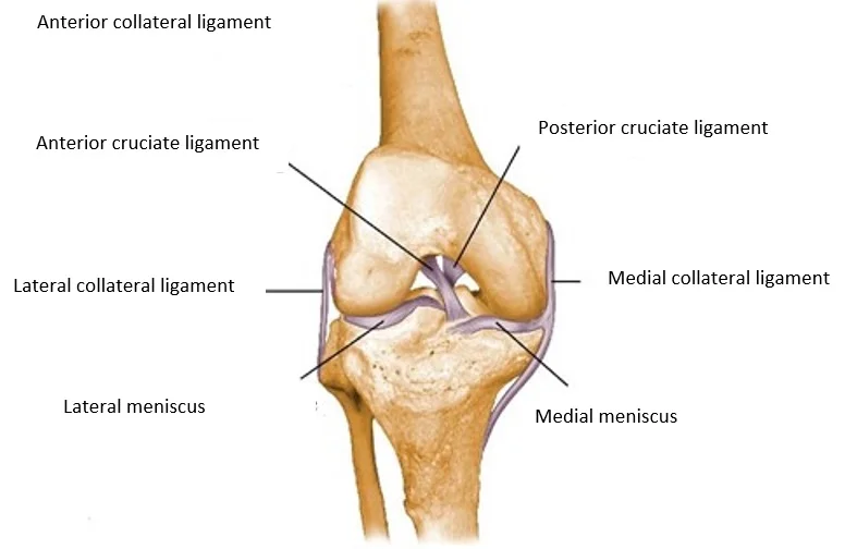
The knee joint has one of the most complex structures in the human body. The structure of the knee joint allows it to contribute to multiple lower limb movements. There are two ring shaped, semilunar cartilages (menisci) between the thighbone and the shinbone. These consist of fibrous cartilage, and they are the shock absorbers of the knee joint. However, those parts of the thighbone, shinbone, and kneecap, which are within the joint, are covered with a different type of cartilage, the so called hyaline cartilage. The concurrent effect of genetics, biomechanics, and lifestyle factors may cause early degenerative changes; these are the so called “tissue wear” changes because of which knee symptoms develop, movement may become painful, so the joint function may be damaged. Additionally, the knee joint may get injured during numerous different movements too, which most often happen during sport, recreational activities, or work, thus the pathology may be further aggravated by them.

General information about the disease
The causes of the symptoms are often the commencing joint wear, the accompanying relative instability, but mostly, the axis deviation of the knee (X leg, O leg). Depending on the load, sometimes the knee is painful, and sometimes it is not, so very often the problem is only detected later due to the fluctuating symptoms, and other concurrent conditions.
Uneven loading may develop due to an incorrect knee joint axis (X leg, O leg, incorrect position of the kneecap), which may result in early joint wear, joint deformities, and ligament failure. A corrective surgery of the knee axis may be performed to prevent the above, or to reduce the pain which is already present.
If the axes are not corrected and the cartilage damages are not treated in time, the biomechanical setting will wear the joint surfaces, which may lead to cartilage wear (arthrosis) over time. The aim of the knee surgery is to make the knee painless, protect the joint surfaces of the knee from potential additional damages, and to prevent cartilage wear, which will occur over time.
Which are the potential symptoms?
Common symptoms affecting the knee joint such as:
- Swelling
- Diffuse pain
- Limited range of motion in the joint
- Obstructions
- Variable knee joint symptoms (alternation of painless and painful periods of time)
- Pain at the outer/inner side of the knee

What are the treatment options?
Basically, there are two types of treatment options available. Conservative (non surgical) and surgical treatment.
The aim of conservative treatment:
The main goal of the treatments is to achieve a painless condition, and to slow down the degenerative changes in the knee. Unfortunately, the area with cartilage loss does not heal either with conservative, or with surgical treatment, so it is expected that the joint wear of the knee will increase with time, which in years, may result in a knee joint that is painful and difficult to use even in everyday life. Loading axes and the work related or sport related loads may vary from patient to patient, so the course of the disease and its velocity varies from person to person too. Last, but not least, body weight influences the achievable results negatively.
Conservative treatment methods
In case of acute knee symptoms, it is necessary to elevate and cool (icing, gel ice packs) the injured limb, and to not put any weight on it. Please do not continue doing sports. Fluid retention may develop in the joint, and the knee may swell up. Pharmacological treatment, using painkillers, and anti inflammatory drugs, physiotherapy, and other methods of physical therapy are all parts of conservative treatment.
Once the acute symptoms are reduced, the administration of cartilage repair agents orally or through injections into the joint, strengthening of the thigh muscles, physiotherapy, and physical therapy may significantly alleviate your symptoms.
Knee surgery
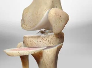
Your surgeon may recommend surgery if significant condition deterioration is expected within 10 years, during which the cartilage damages are treated with a minimally invasive arthroscopic procedure through small incisions, then the loading axes are corrected from a surgical exploration by cutting through the shinbone. The aim of the arthroscopic interventions is to remove the damaged cartilage piece by sparing as much healthy cartilage as possible. After the arthroscopic surgery, metal fixation is used for the maintenance of the new axis, which may be removed on a later occasion based on individual evaluation, after the bone has healed. Recovery heavily depends on the surgical findings, concurrent injuries, and the postoperative treatment which is supported by physiotherapists.
Antibiotic and pathological coagulation prevention is used during surgery, if necessary, which should be continued at home in some cases. Additionally, it is necessary to perform the practiced physiotherapy exercises, and to rest the operated limb at home in accordance with the instructions of the physician, who performed the surgery.
It is necessary to refrain from putting any weight on the limb by using crutches for 6 weeks after the intervention. In non problematic cases, the patients are discharged home on the first day after surgery. Anticoagulation should be applied for a determined period of time after surgery, which could be administered by the patient too.
The surgical process
During the surgery, two to three incisions of 1 cm are made on both sides of the knee, so that the arthroscope and the arthroscopic devices could be inserted into the knee joint.
During the intervention, any potential damaged parts of the knee cartilage are removed through the tiny hole with devices, which are small in size, and the cartilage surface injuries are smoothed out.
The area is rinsed, so that the inflammatory fluids and debris are removed.
Following this, axis correction, and fixation of the bones in their new position are performed from a new skin incision on the outer or inner side of the knee (depending on the applied technique); pieces of medical alloys are used for the fixation. A small drain is inserted into the wound, if necessary, which will remove the blood and plasma that will accumulate after the surgery.
Elastic bandage is applied on the operated lower limb after the surgery.
What happens if the justified surgical treatment is not performed?
The damaging of the cartilage surfaces may accelerate, making the pain and the swelling in the knee joint more intense. The damaging of the joint’s cartilage surface may continue, which may lead to joint wear (arthrosis) in the long term. This process is irreversible, and it may result in a very painful joint with reduced range of motion, which could only be managed by additional knee surgery.
When can you return to your everyday activities?
Sedentary work usually can be started 3 weeks after the knee surgery, but physical labor can only be started 3 months after the intervention. When it comes to sports, using a stationary bike can be started after 6 weeks; swimming can be started after 6 weeks and jogging after 3 months provided that the wound has healed. In order to achieve full recovery, other sports (football, basketball, etc.), and skiing can only be started half a year later.
If you have any questions, please send a letter to magankorhaz@bhc.hu!


