2022-12-08 16:15 || 1.0.0
  • Szolgáltatásaink
  • Árlista
  • Kórházfejlesztés
  • Magunkról
  • Kapcsolat
  • Karrier
  • Telefon: (+36) 1 489-52-00 | E-mail: info@bhc.hu

    Ultrasound

    Az ultrahang egy olyan képalkotó technika, mely során az emberi fül számára nem hallható hanghullámok segítségével kap a vizsgáló orvos vizuális információt kép formájában. Az eljárás fájdalommentes és a szervezetet nem károsítja.

    Az ultrahangos vizsgálatok előnyei

    Az eljárás az orvostudomány jelenlegi állása szerint nem jár kockázattal szemben a CT vizsgálatokkal és röntgennel, melyek ionizáló sugarakat használnak. Az ultrahangot a medicina számos területe alkalmazza, például a belgyógyászat, kardiológia, az endokrinológia, a nőgyógyászat, érsebészet, urológia és az ortopédia. A módszer valós időben ad képet, így akár mozgás közben is megfigyelhető a vizsgált szerv.

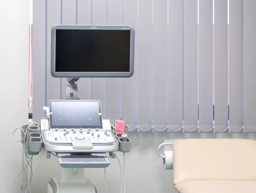
    Ultrasound

    Limitations of ultrasound examinations

    Ultrasound travels poorly in air and does not pass through bones, so it is not suitable for examining organs that are filled with air or covered by bones, such as the lungs or the brain. In the deeper layers of the body, the examination may be limited.

    How are ultrasound examinations carried out?

    Most of the time, ultrasound examinations do not require any preparation, but there are exceptions. For example, abdominal examinations should be performed on an empty stomach, and pelvic examinations should be performed optimally with a full bladder. The area studied by ultrasound must be exposed, for this it may be necessary to remove jewelry and pull aside clothing. The proper propagation of ultrasound is facilitated by a gel that can be easily wiped off and washed off the skin. In some cases, the ultrasound recording is not performed through the skin, but the doctor performs a body cavity examination.